Melasma, freckles and age spots are all forms of hyperpigmentation (dark spots), but they are fundamentally different — and the fastest way to tell them apart is by four clues: shape, location, age of onset and how they change with the seasons. Melasma appears as diffuse, symmetric patches on the cheeks, forehead and upper lip, driven by hormones and UV, chronic and prone to relapse; freckles are small, light-brown macules with a genetic basis that appear in childhood and fade in winter; solar lentigines (age spots / sun spots) are larger, well-defined spots caused by cumulative sun damage that do not fade seasonally; and post-inflammatory hyperpigmentation (PIH) is a dark mark that appears exactly where acne or an injury once was. Identifying the correct type is a mandatory first step, because each one needs a different treatment approach — and treating the wrong one (especially aggressive laser for melasma) can make things worse, not better.
This article is a practical guide to distinguishing the pigmentation types most often confused on Vietnamese skin: melasma (epidermal/superficial, dermal/deep, and mixed), freckles, solar lentigines and post-acne marks. You will get a detailed comparison table, an at-home identification guide, and a clear explanation of why "diagnose first, treat second" matters so much. Content is compiled from the American Academy of Dermatology (AAD), DermNet NZ, StatPearls (NCBI) and review articles on the US National Library of Medicine (PMC), combined with real clinical experience at Eternal Beauty Center — a skincare clinic in Go Vap, Ho Chi Minh City. To go deeper on melasma alone, read our overview: Melasma: causes and treatment.
Quick look: four "dark spots" people mix up
Not every dark spot on the face is melasma. On Vietnamese skin, four pigmentation conditions are commonly lumped together as "melasma" or "freckles" when they are really four different problems: melasma, freckles (ephelides), age spots / solar lentigines, and post-inflammatory hyperpigmentation (PIH). They differ in cause, in how deep the pigment sits in the skin, and — most importantly — in how they respond to treatment.
In simple terms, every dark spot is the result of excess melanin produced and deposited in the skin. But where the melanin sits (superficial epidermis vs deeper dermis), what triggers it (hormones, genetics, sun, or inflammation) and how it is distributed (scattered spots vs diffuse patches) determine which type it is. That is exactly why an over-the-counter "anti-melasma" product can do nothing for one person yet work for another — because they actually have two different conditions.
Three questions point you in the right direction from the start:
Is it scattered spots or a diffuse patch? Small separate spots suggest freckles/age spots; large symmetric patches suggest melasma.
When did it appear, and does it change with the seasons? Present since childhood and fading in winter → freckles; appearing in middle age and staying put → age spots.
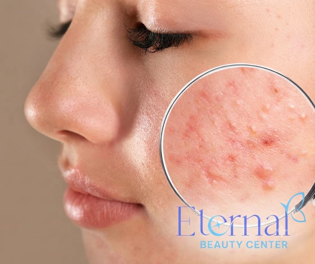
Does it sit exactly where acne or an injury once was? If it tracks an old blemish → post-inflammatory hyperpigmentation.
The next four sections describe each type, followed by a summary comparison table.
What is melasma — and its 3 depth types
Melasma is an acquired form of hyperpigmentation that shows up as brown to grey-brown patches distributed symmetrically on both sides of the face — most often on the cheeks, forehead, bridge of the nose, upper lip and chin. Per DermNet NZ and the AAD, melasma is driven mainly by two combined factors: hormones (estrogen/progesterone) and ultraviolet (UV) light, which is why it is especially common in women of reproductive age, during pregnancy (the "mask of pregnancy" — chloasma) or when using hormonal medication. Heat and visible light (including blue light from screens) can also trigger it.
Two key diagnostic features of melasma: symmetry (it appears almost identically on both cheeks) and ill-defined, diffuse patch-like borders — completely different from the scattered, sharply bordered spots of freckles or age spots. Melasma also typically spares the area around the eyes. One thing to accept up front: melasma is a chronic, relapsing condition — meaning it can be controlled, lightened and maintained, but there is no way to "cure it once and for all permanently." Any promise of "100% melasma removal" should make you cautious.
The factor that decides treatment is the depth of the pigment. The literature divides melasma into three types:
Epidermal melasma (superficial / patch melasma). Pigment sits mainly in the epidermis, is a clear brown, with relatively defined borders. Under a Wood's lamp, the affected area appears darker and more enhanced — a sign the pigment is superficial. This type responds best to topical actives and peels.
Dermal melasma (deep melasma). Pigment sits deep in the dermis, appears grey-brown or blue-grey, with blurred borders. Under a Wood's lamp it is less enhanced because the melanin is deep. This is the hardest type to treat, responds slowly, and relapses easily.
Mixed melasma. A combination of both — the superficial part responds faster, the deep part slowly. This is the most common type in clinical practice.
So the question "is it superficial or deep melasma" is not semantics — it completely changes the expectations and the protocol. Classifying depth requires a dermatologist's assessment using examination, a Wood's lamp or a dermatoscope; it cannot be guessed reliably from a photo.
What are freckles (ephelides)
Freckles (ephelides) are small, flat, light-brown to brown macules, 1–3mm in size, usually clustered on sun-exposed areas such as the cheeks, bridge of the nose, shoulders and backs of the hands. Per DermNet NZ and StatPearls, freckles have a clear genetic basis (linked to the MC1R gene), are common in fair-skinned people with blond/red hair, and typically appear in early childhood (from about age 2–3), darkening through the teenage years.
The most distinctive clue for freckles is seasonal change: they darken in summer with more sun and fade noticeably in winter or when sun is carefully avoided. This is a major difference from age spots (which don't fade seasonally) and melasma (which doesn't fluctuate this way). Fundamentally, freckles arise because pigment cells (melanocytes) produce more melanin when exposed to UV, not because the number of pigment cells increases.
Freckles are benign and, for many people, a charming feature. If you want to fade them, targeted pigment methods (such as selective laser, light-based therapy or brightening actives) can be effective — but sun protection remains the foundation, because without it freckles return every summer. Worth remembering: freckles often respond to laser/light better than melasma, which is one reason you must never treat "melasma and freckles" with a single blanket protocol.
What are age spots / solar lentigines
Age spots (solar lentigo, also called sun spots or liver spots) are flat brown spots with well-defined borders, larger than freckles (a few mm to over 1cm), caused by cumulative sun damage over time. Per DermNet NZ, solar lentigines appear mainly from middle age onward in areas exposed to years of sun: the face, backs of the hands, forearms and shoulders. Unlike freckles, age spots form from a mild increase in the number of pigment cells in the basal layer plus increased melanin production, so they are persistent and do not fade seasonally — once they appear they stay year-round, may darken, but essentially do not disappear on their own.
Identifying features: separate spots, sharply defined borders, uniform color, flat with the skin surface. They are usually few in number and increase with age and a lifetime of sun exposure. Because age spots reflect years of sun damage, they are also a reminder of sun-related skin risk — so any spot that changes rapidly in color, size, border, or bleeds should be checked by a dermatologist to rule out a malignant lesion.
For treatment, because the pigment is localized with clear borders, age spots often respond well to targeted pigment methods such as selective laser, intense pulsed light (IPL) or cryotherapy — unlike melasma, which needs a gentle, comprehensive approach. This is why distinguishing "is this an age spot or melasma" directly affects whether laser should be used at all.
What is post-inflammatory hyperpigmentation (PIH)
Post-inflammatory hyperpigmentation (PIH) is a dark mark that appears exactly where the skin was previously inflamed or injured — after acne, a wound, a burn, eczema, or after skin procedures. The mechanism: the inflammatory process stimulates pigment cells to produce more melanin, or causes melanin to "drop" into the deeper dermis. This is a very common type of hyperpigmentation in people with darker skin (typical of Vietnamese skin tones), and in hot, humid climates the marks are often darker and slower to fade.
Key distinguishing clue: PIH tracks an old mark — you can usually recall "this is where I had a pimple/injury." The mark is flat with the skin surface (unlike an atrophic scar, which is depressed) and is purely a change in color. The good news is that PIH can fade on its own over months or years if the root cause is controlled (acne clears, inflammation stops) and sun protection is strict; the process can be accelerated with tyrosinase-inhibiting actives and appropriate peels.
Because PIH starts from inflammation, rule number one is to address the cause first — for example, fully treating the acne — before working on the leftover "color." If you only focus on brightening the marks while the acne keeps coming back, new marks will keep appearing. Our article Why do dark marks linger after a pimple heals? explains this mechanism and how to handle it.
Detailed comparison table: identify it in 1 minute
The table below summarizes the key distinguishing features of the four pigmentation types. It is a quick identification tool — but the final diagnosis, especially determining melasma depth, still needs a dermatologist.
| Criterion | Melasma | Freckles (ephelides) | Age spots (solar lentigo) | Post-acne marks (PIH) |
|---|---|---|---|---|
| Shape | Diffuse patch, blurred border, symmetric | Small 1–3mm scattered spots | Larger spots (mm–cm), sharp border, flat | Mark/spot tracking an old injury |
| Color | Brown to grey-brown (deep: blue-grey) | Light brown to brown | Uniform brown | Brown to dark brown/purple |
| Main cause | Hormones + UV (+ heat, light) | Genetics (MC1R) + UV | Cumulative sun damage + age | Prior inflammation/injury |
| Typical location | Cheeks, forehead, upper lip, chin | Cheeks, nose, shoulders, hands | Face, hands, forearms, shoulders | Exactly where acne/injury was |
| Age of onset | Reproductive age (20–40+), pregnancy | Childhood, darker in teens | Middle age onward | Any age, after skin inflammation |
| Seasonal change | Little; worse with sun/heat | Darker in summer, fades in winter | Does not fade seasonally | No; fades gradually over time |
| Pigment depth | Superficial / deep / mixed | Superficial (epidermal) | Epidermal (basal layer) | Epidermal and/or dermal |
| Wood's lamp | Superficial: enhanced · Deep: less so | Enhanced | Enhanced | Depends on depth |
| Treatment approach | Gentle, multi-active + strict sun protection; laser cautiously | Sun protection + selective laser/light | Targeted laser/IPL/cryotherapy | Treat the cause first + brighten/peel |
| Difficulty & outlook | Hardest; chronic, relapsing | Easier; returns without sun protection | Responds well to targeted procedures | Moderate; fades if inflammation stops |
If you remember only one thing from this table: melasma responds very differently to laser than the others. The next section explains why.
Why the right diagnosis decides the treatment
Identifying the correct pigmentation type matters because each responds differently — and with melasma, the wrong treatment (especially aggressive laser) can make it worse rather than better. This is the core message of the whole article.
Melasma needs a gentle, patient approach. The pigment cells in melasma are overactive and easily provoked; any inflammatory or heat trigger — including laser set too aggressively — can make melanocytes "backfire." The literature clearly documents rebound hyperpigmentation after laser in melasma patients, especially in darker skin: in some studies melasma recurred in all patients, and sometimes came back darker than before treatment. This is why the AAD emphasizes that the foundation of melasma care is strict sun protection + gentle topical actives (tyrosinase inhibitors), and that most patients should not start with laser. If energy is used, it must be conservative parameters, applied cautiously and by experienced hands.
By contrast, freckles and age spots usually respond well to targeted pigment methods (selective laser, IPL, cryotherapy), because the pigment is localized, the borders are clear, and the melanocytes are not as "easily angered" as in melasma. Here lies the dangerous trap: if you mistake melasma for an age spot and treat it with aggressive laser, you risk flaring the melasma. Conversely, if you mistake an age spot for melasma and only apply brightening cream, the age spot may fade very slowly or barely at all.
Post-acne marks must have the underlying inflammation addressed first. If you only chase the marks while acne keeps recurring, new marks keep forming — wasted effort. And if you mistake melasma for post-acne marks, people tend to reach for actives/peels that are too harsh, causing irritation that can inadvertently ignite melasma.
In short, a wrong diagnosis is not only less effective but can sometimes cause real harm. That is why at Eternal Beauty Center the principle is always to examine, classify and grade first, then design the protocol. And because UV is the common denominator of all four types, correct sun protection is a non-negotiable foundation regardless of your type.
How to tell them apart at home and when to see a dermatologist
You can get an initial sense at home with a few simple observations — although an accurate diagnosis (especially melasma depth) still needs a doctor. Look at your face in natural light and answer:
1. Symmetric or not? Brown patches appearing almost identically on both cheeks/forehead → think melasma. Spots in just one area, scattered → lean toward freckles/age spots. 2. Scattered spots or a diffuse patch? Many small separate spots → freckles (if small, present since childhood) or age spots (if larger, sharply bordered, middle age). A large patch with blurred edges → melasma. 3. When did it appear and does it change seasonally? Present since childhood, darker in summer, fading in winter → freckles. Appearing in middle age, fixed year-round → age spots. 4. Does it sit exactly where acne/an injury was? If the dark mark is right where you had a pimple → post-acne marks. 5. Is it raised or depressed to the touch? All four types of hyperpigmentation are flat, only a color change. If the surface is bumpy, scaly, or depressed → have a doctor assess it (it may be something other than simple pigmentation).
See a dermatologist when:
You are not sure whether it is melasma, an age spot or post-acne marks — because the treatment paths differ completely.
You suspect melasma and want to determine superficial/deep/mixed to choose the right protocol and set realistic expectations.
A spot changes rapidly in color, size or shape, has an irregular border, bleeds or itches — a malignant lesion must be ruled out.
You have used a brightening product for several months with no improvement, or your skin became irritated or darker after using it.
You have had laser and ended up darker — a classic sign of rebound hyperpigmentation that needs re-evaluation.
Getting examined early saves you a costly detour: treating the right type from the start is always faster and safer than fixing a mistake later.
Treating each type correctly at Eternal Beauty Center
At Eternal Beauty Center (Go Vap, HCMC), hyperpigmentation is handled exactly in the spirit of this article: diagnose and classify first, personalize the protocol second — not "one treatment for everyone." A dermatologist (Dr. Le Hien) personally examines the skin, distinguishes melasma from freckles/age spots/marks, and for melasma classifies it as superficial, deep or mixed to set realistic expectations before starting.
Depending on the diagnosis, the following directions can be selected and combined:
E-Mela — non-invasive melasma protocol: for confirmed melasma, especially deep and mixed melasma. Built by Dr. Hien Le, E-Mela uses a new-generation Resorcinol (a tyrosinase inhibitor) that targets pigment sitting deep (over 200 cell layers), with no peeling, no ablation and no invasion — consistent with the "gentle for melasma" principle the literature recommends. The protocol is personalized after the dermatologist examines and classifies the melasma.
Mela Peel — specialized pigment peel: a peel designed specifically for pigmentation, suited to superficial melasma and post-acne marks (PIH) — addressing the leftover "color" in the superficial layer in a controlled way.
Eternal's core difference is the correct order: diagnosis/classification first — precisely the message of this article. Because melasma is chronic, we do not promise "permanent removal"; instead, the realistic goal is to lighten, control and maintain, with strict sun protection to limit relapse. After the skin exam, the doctor gives an honest forecast of expected improvement and the timeline needed (results vary from person to person).
Frequently asked questions (FAQ)
How do I tell melasma from freckles?
Melasma appears as diffuse patches with blurred borders distributed symmetrically on the cheeks, forehead and upper lip, driven by hormones and UV, usually appearing in reproductive age. Freckles are small scattered spots (1–3mm), light brown, with a genetic basis, appearing in childhood and — most tellingly — darkening in summer and fading in winter. In short: melasma is a "patch" that doesn't fade seasonally, while freckles are "spots" that clearly change with the seasons.
What is the difference between deep melasma and epidermal (patch) melasma?
The difference is pigment depth. Epidermal (superficial/patch) melasma has melanin in the epidermis, a clear brown color, relatively defined borders, appears more enhanced under a Wood's lamp, and responds well to topical actives and peels. Deep (dermal) melasma has melanin deep in the dermis, a grey-brown or blue-grey color, blurred borders, is less enhanced under a Wood's lamp, and is much harder to treat, responding slowly and relapsing easily. Many people have mixed melasma — a combination of both.
Is a dark spot on my face melasma or an age spot?
Age spots (sun spots, liver spots) are scattered brown spots with sharp borders, flat, larger than freckles, appearing in middle age from cumulative sun damage, and they do not fade seasonally. Melasma is a diffuse patch with blurred borders, symmetric, linked to hormones. If they are separate spots with clear borders in long-sun-exposed areas, they are more likely age spots; if it is a symmetric patch on both cheeks, it is more likely melasma. An accurate diagnosis should be confirmed by a dermatologist.
Why does it matter to identify the correct pigmentation type?
Because each type responds differently to treatment. Melasma needs a gentle, multi-active approach with strict sun protection and reacts badly to aggressive laser (which can cause rebound hyperpigmentation and make it darker). Freckles and age spots, by contrast, usually respond well to selective laser/light, while post-acne marks require addressing the underlying inflammation first. A wrong diagnosis leads to the wrong treatment — ineffective, and sometimes making the condition worse.
Is laser treatment for melasma safe?
Laser is not a first-line choice for melasma. The pigment cells in melasma are easily provoked, so laser set too aggressively can cause rebound hyperpigmentation and make melasma relapse, sometimes darker than before, especially in darker skin such as Vietnamese skin. Per the AAD, the foundation of melasma care is strict sun protection combined with gentle topical actives; if energy is used, it must be conservative parameters, applied cautiously by a qualified professional after the melasma has been classified.
Do freckles and age spots go away on their own?
Freckles don't disappear completely but fade noticeably in winter or with careful sun avoidance, and darken again in summer. Age spots are persistent, do not fade seasonally, and essentially do not go away on their own because they reflect cumulative sun damage. Both are benign, but any spot that changes rapidly in color, size or border should be checked by a dermatologist to rule out a malignant lesion.
How is post-acne hyperpigmentation different from melasma?
Post-acne marks (PIH) are dark marks that appear exactly where the skin was previously affected by acne or inflammation/injury, and they can fade on their own over months or years if the cause is controlled and sun protection is strict. Melasma is a diffuse, symmetric patch caused by hormones and UV, chronic and prone to relapse. The distinction: marks track an old lesion and tend to fade, while melasma appears as symmetric patches and is more persistent.
Can melasma be permanently cured?
Melasma is a chronic, relapsing condition, so the realistic goal is to lighten, control and maintain rather than "cure it once and for all permanently." With the correct type identified, a gentle protocol matched to the depth, and strict sun protection, most cases can improve noticeably and stay stable. Be cautious of any promise of "100% melasma removal."
Summary
Melasma, freckles, age spots and post-acne marks are all hyperpigmentation but four different problems — and identifying the right one is a mandatory step to treating it correctly. The most important takeaways:
Melasma = brown/grey-brown patches, symmetric, from hormones + UV, chronic and relapsing; three depth types (patch/superficial – deep – mixed), and depth decides the protocol.
Freckles = small spots, genetic, present since childhood, darker in summer, faded in winter.
Age spots = larger spots, sharp borders, from cumulative sun + age, fixed year-round.
Post-acne marks = dark marks tracking an old injury, fade gradually once inflammation stops.
The right diagnosis changes the treatment: melasma needs gentle care + strict sun protection and avoids aggressive laser (rebound risk); freckles/age spots respond well to selective laser; post-acne marks need the inflammation addressed first.
Because UV is the common denominator, sun protection is the foundation for every type. And because a wrong diagnosis can cause harm, let a dermatologist examine and classify your skin before choosing a method — rather than guessing from a photo or buying products off an ad.
Book a skin exam & pigmentation-classification consult at Eternal Beauty Center
Unsure whether the dark spots on your face are melasma, freckles, age spots or post-acne marks? A dermatologist at Eternal Beauty Center will examine, distinguish and accurately classify them (including grading melasma as superficial or deep), then advise a personalized protocol with an honest forecast of the timeline.
Hotline / Zalo: 0334 713 610
Address: 204 Street No. 1, Ward 16, Go Vap District, Ho Chi Minh City
Opening hours: 10:00 – 20:00 daily
Explore services: E-Mela – Non-invasive melasma treatment · Mela Peel – Pigment & marks peel
Read more: Melasma: causes & treatment · Sun protection done right · Why you get post-acne marks
Medical disclaimer: This article is for informational purposes and does not replace an in-person examination, diagnosis or individual medical advice. Distinguishing and treating hyperpigmentation should be assessed by a qualified physician; effectiveness and suitability vary from person to person depending on the lesion type, pigment depth and individual skin. If a pigmented spot changes rapidly in color, size or border, see a dermatologist to rule out a malignant lesion. Please consult a physician before starting any protocol.